

I. Introduction
II. Experimental Methods
2.1 Materials and image acquisition
2.2 Image Analysis
III. Results
3.1 Quantitative comparison of ACHI and CNR according to coupling material
3.2 Inferential statistical analysis of ACHI and CNR
3.3 Near field artifact burden quantified by HEAF
IV. Discussion
V. Conclusion
I. Introduction
Acoustic coupling at the transducer tissue interface is essential for superficial ultrasound imaging because small air gaps and impedance mismatch can degrade near field signal transmission and image fidelity.[1,2] Stand off pads increase the distance between the transducer and superficial target structures, allowing the region of interest to be positioned within a more appropriate focal zone and improving near field visualization.[3,4] However, conventional gel based media may be displaced by probe pressure, may not maintain stable contact on irregular surfaces, and may generate acoustic shadowing or near field artifacts when air bubbles are present at the gel skin interface.[2,3,4] To overcome these limitations, pad type coupling materials such as hydrogel pads, stand off pads, and bolus type materials have been proposed.[3,4,5,6] Previous studies on pad type coupling materials have primarily evaluated image quality using signal- or contrast-based metrics such as Sound to Noise Ratio (SNR) and Contrast-to-Noise Ratio (CNR) under specific disease conditions, anatomical sites, or single material comparisons.[5,6,7,8] However, near field high echo artifacts arising at the coupling interface are a clinically important but underexplored issue: they can obscure superficial structures such as skin nodules, tendons, and superficial vessels, impair lesion characterization, and interfere with Doppler blood flow assessment.[6,7] No prior study has systematically quantified near field artifact generation across different pad type materials under identical imaging conditions. For quantitative assessment, the classical SNR in fully developed Rayleigh speckle is known to converge to approximately 1.91.[9] However, ex vivo tissue images contain structural echoes from muscle fibers, fat layers, and connective tissue; therefore, the μ/σ ratio was defined in this study as the Acoustic Coupling Homogeneity Index (ACHI), rather than as a strict speckle SNR. Unlike ACHI and CNR, which reflect signal homogeneity and contrast within predefined Regions Of Interest (ROIs), High-Echo Artifact Fraction (HEAF) was designed to quantify near field high echo artifacts generated specifically at the coupling interface, providing complementary information on artifact burden that cannot be captured by conventional signal- or contrast-based metrics alone. This study aimed to compare HydroAid®, Aquaflex®, and Bolx-II® with conventional gel using ACHI, CNR, and HEAF, and to evaluate their applicability as pad type coupling materials for superficial ultrasound imaging.
II. Experimental Methods
2.1 Materials and image acquisition
Three commercially available pad type acoustic couplants were evaluated: HydroAid® hydrogel dressing, Aquaflex® ultrasound gel pad, and Bolx-II® radiotherapy bolus material. Conventional gel was included as a reference condition. The physical and acoustic properties of the four coupling materials are summarized in Table 1. For pad type materials, a 1 cm stand off layer was placed between the transducer and the tissue surface, whereas gel was applied directly to remove air gaps and maintain acoustic coupling. ACHI, CNR, and HEAF were compared across all four coupling conditions. The approximate acoustic impedance values of the materials ranged from 1.48 MRayl to 1.58 MRayl, which is close to the soft tissue range. Therefore, acoustic impedance mismatch alone is unlikely to be the primary determinant of the observed differences in image quality metrics. However, attenuation coefficients were not directly measured in this study; therefore, the contribution of material specific attenuation to ACHI and CNR differences should be evaluated in future studies.
Table 1.
Physical and acoustic properties of the coupling materials used in this study.
2.2 Image Analysis
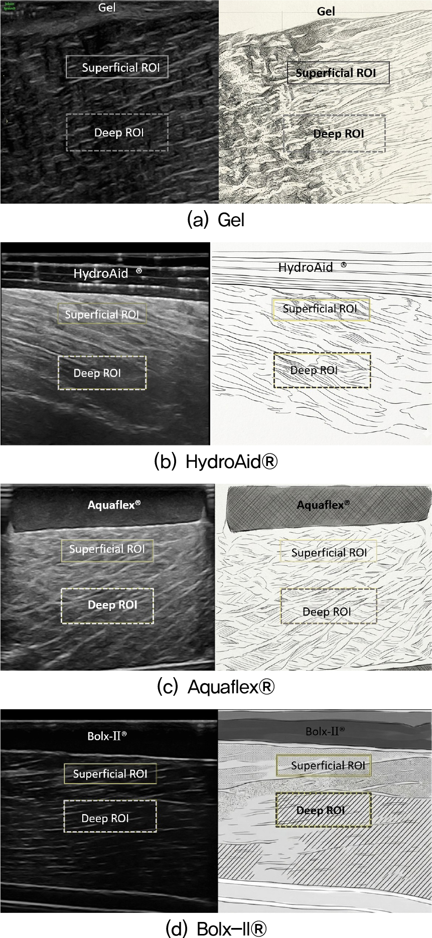
All acquired B mode ultrasound images were analyzed using Fiji (ImageJ, NIH, Bethesda, MD, USA). As shown in Fig. 1, two ROIs were defined in each image: a superficial ROI within the hyperechoic layer immediately beneath the coupling interface (462 pixel × 98 pixel) and a deep ROI within the muscular tissue layer (460 pixel × 152 pixel). ROI boundaries were positioned to exclude reflection lines, structural discontinuities, and acoustic shadowing.
Mean pixel intensity (μ) and standard deviation (σ) were extracted from each ROI and used to calculate three quantitative indices. The ACHI was defined as μ/σ, sharing the same formulation as conventional SNR but interpreted as a relative homogeneity index rather than a strict Rayleigh speckle SNR, given that ex vivo tissue ROIs inherently contain structural echoes from muscle and fat.[9] Superficial and deep ACHI values were defined as Eq. (1):
The CNR was calculated by normalizing the absolute difference in mean pixel intensity between the superficial and deep ROIs by their combined standard deviation,as given in Eq. (2):[10,11]
The HEAF was defined as the proportion of pixels within the near field region immediately beneath the coupling interface that exceeded an intensity threshold of μ_S + σ_S.[12] Pixels above this level were classified as high-echo artifact pixels attributable to near field reverberation and backscattering,as given in Eq. (3):
where N_high is the number of pixels exceeding the threshold and N_total is the total pixel count in the near field ROI.
III. Results
3.1 Quantitative comparison of ACHI and CNR according to coupling material
Quantitative image quality metrics according to coupling material are summarized in Table 2. Across all three ex vivo tissue samples, HydroAid® and Aquaflex® generally showed higher ACHI and CNR values than Bolx-II®.
Table 2.
Comparison of ACHI and CNR according to coupling material in three tissue samples (mean ± SD, n = 10).†
| Phantom | Metric | Gel | HydroAid® | Aquaflex® | Bolx-II® |
| Beef rump | 1.969 ± 0.390 | 4.682 ± 0.850 | 5.172 ± 0.921 | 1.744 ± 0.334 | |
| 2.011 ± 0.605 | 3.092 ± 0.566 | 2.956 ± 0.364 | 1.756 ± 0.324 | ||
| 0.458 ± 0.374 | 1.061 ± 0.791 | 1.826 ± 0.768 | 0.345 ± 0.259 | ||
| Pork loin | 1.747 ± 0.211 | 5.055 ± 0.823 | 5.721 ± 0.881 | 1.402 ± 0.230 | |
| 2.883 ± 0.775 | 4.104 ± 0.908 | 4.449 ± 0.488 | 1.528 ± 0.156 | ||
| 0.943 ± 0.565 | 1.063 ± 0.456 | 0.748 ± 0.363 | 0.293 ± 0.255 | ||
|
Pork tenderloin | 1.629 ± 0.304 | 6.192 ± 1.196 | 5.162 ± 0.542 | 1.732 ± 0.167 | |
| 1.938 ± 0.473 | 3.936 ± 0.551‡ | 3.032 ± 0.301 | 1.636 ± 0.126 | ||
| 0.421 ± 0.366 | 1.249 ± 0.486 | 1.386 ± 0.237 | 0.017 ± 0.163 |
In the beef rump sample, Aquaflex® showed the highest superficial ACHI (ACHI_S, 5.172 ± 0.921) and CNR (1.826 ± 0.768), whereas HydroAid® showed the highest deep ACHI (ACHI_D, 3.092 ± 0.566). In the pork loin sample, Aquaflex® showed the highest ACHI_S and ACHI_D values (5.721 ± 0.881 and 4.449 ± 0.488, respectively), while HydroAid® showed the highest CNR (1.063 ± 0.456). In the pork tenderloin sample, HydroAid® showed the highest ACHI_S and ACHI_D values (6.192 ± 1.196 and 3.936 ± 0.551, respectively), whereas Aquaflex® showed the highest CNR (1.386 ± 0.237).
Overall, HydroAid® and Aquaflex® demonstrated comparable image quality metrics, while Bolx-II® consistently showed the lowest or near lowest ACHI and CNR values across the tissue samples.
3.2 Inferential statistical analysis of ACHI and CNR
Inferential statistical analysis revealed significant differences in ACHI_S, ACHI_D, and CNR among coupling materials across all three tissue samples (all p < 0.001; Table 2). Post hoc analysis showed that HydroAid® and Aquaflex® consistently demonstrated significantly higher ACHI and CNR values than Bolx-II®, whereas differences between HydroAid® and Aquaflex® were generally not significant. The only exception was ACHI_D in the pork tenderloin sample, where HydroAid® was significantly higher than Aquaflex®.
3.3 Near field artifact burden quantified by HEAF
HEAF values for the four coupling conditions are summarized in Table 3 and Fig. 2. HEAF varied according to both coupling material and tissue type. Conventional gel showed nearly zero HEAF in all tissue samples, whereas the pad type materials showed higher values to varying degrees. Because HEAF values were concentrated near zero in several groups, differences among coupling materials were analyzed using the Kruskal–Wallis test followed by Dunn–Bonferroni post hoc comparisons.
Table 3.
HEAF by coupling condition and tissue sample (mean ± SD, n = 10).
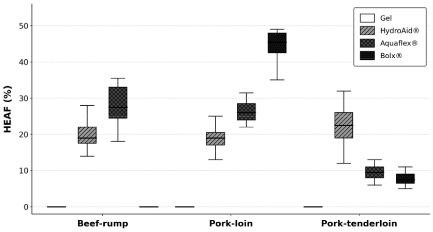
HEAF differed significantly among coupling materials in all three tissue samples: beef rump (H = 35.11, p < 0.001), pork loin (H = 35.56, p < 0.001), and pork tenderloin (H = 34.85, p < 0.001). Among the pad-type materials, HydroAid® consistently showed the highest HEAF in all tissue samples: 28.15 % ± 4.38 % in beef rump, 42.55 % ± 6.77 % in pork loin, and 20.47 % ± 6.67 % in pork tenderloin. Aquaflex® showed intermediate HEAF values, whereas Bolx-II® remained relatively low and close to conventional gel.
Post hoc analysis showed that Aquaflex® and HydroAid® generally had significantly higher HEAF values than conventional gel, while Bolx-II® did not differ significantly from conventional gel in most comparisons. The overall HEAF ranking was as follows: conventional gel ≈ Bolx-II® < Aquaflex® < HydroAid®. These findings indicate that HydroAid® generated the greatest near field high echo artifact burden, whereas conventional gel and Bolx-II® produced minimal near field artifacts under the present imaging and threshold conditions.
IV. Discussion
This study quantitatively compared the acoustic coupling performance and near field artifact characteristics of four pad type ultrasound coupling materials using ex vivo tissue models. HydroAid® and Aquaflex® generally showed higher ACHI and CNR values than Bolx-II®, suggesting that hydrogel- or gel-pad-type materials provide more stable acoustic coupling than a radiotherapy bolus material under near field imaging conditions.
The higher ACHI and CNR values of HydroAid® and Aquaflex® may reflect their flexible gel structures, high water content, and better surface conformity, which can reduce air gaps and stabilize acoustic energy transmission. Previous studies have reported that gel stand off pads and hydrogel based couplants can improve superficial visualization and reduce near field interference.[6,10] In contrast, the lower ACHI and CNR of Bolx-II® may reflect differences in surface adaptability, internal attenuation, or microscopic scattering. Because all materials had similar soft tissue like acoustic impedance values, impedance mismatch alone is unlikely to explain the observed differences.
HEAF showed a distinct pattern from ACHI and CNR. Conventional gel and Bolx-II® produced near zero HEAF, whereas HydroAid® showed the highest values, followed by Aquaflex®. This dissociation indicates that coupling efficiency and near field artifact generation are independent material properties. The elevated HEAF of HydroAid® may be related to its hydrogel network microstructure, which may promote reverberation or backscattering at the coupling interface. Aquaflex® showed relatively high ACHI and CNR with lower HEAF, suggesting a more favorable balance between image quality and artifact suppression.
Clinically, near field high echo artifacts may obscure superficial structures such as skin nodules, superficial vessels, or tendons, and may interfere with Doppler flow assessment.[6] Therefore, HEAF should be considered alongside signal- and contrast-based metrics when selecting coupling materials for superficial ultrasound examinations.
This study has several limitations. Ex vivo tissue models were used instead of standardized phantoms, and tissue specific structures may have influenced ACHI and CNR values. ROI placement and the relative HEAF threshold may introduce measurement variability depending on tissue echogenicity. Repeated measurements from the same scan location may not guarantee full frame independence, and inter rater reliability was not assessed. Future studies should incorporate standardized near field phantoms, evaluate stand off thickness effects, and include multi rater analysis.
Despite these limitations, Aquaflex® showed the most balanced performance between image quality and artifact suppression, suggesting its potential as a pad type coupling material for superficial ultrasound examinations.
V. Conclusion
HydroAid® and Aquaflex® demonstrated superior ACHI and contrast performance compared with Bolx-II® across all ex vivo tissue models. However, HydroAid® produced the highest HEAF, indicating greater near field artifact generation at the coupling interface. Aquaflex® maintained comparable image quality with lower artifact burden, representing the most balanced performance among the pad type materials evaluated. These findings support the inclusion of HEAF as a complementary metric alongside ACHI and CNR for selecting coupling materials in superficial ultrasound examinations where near field artifact suppression is clinically relevant.
